The aim of our research is to better understand the dynamics of the immune system in the context of human clinical disease. The main focus is to directly visualize the behavior of immune cells during inflammation, infection and resolution by using cutting edge technology, including laser-scanning confocal and 2-photon microscopy. Imaging complex cellular behaviors in real time, both in vitro and in vivo, provides a unique window into these dynamic processes (Jorch & Deppermann, Front Cell Dev Biol., 2021). We focus on monocytes, macrophages and neutrophils in different organs during S. aureus and E. coli infections or diseases like Familial Mediterranean Fever.
Autoinflammatory syndromes including Familial Mediterranean Fever (FMF), PAPA-Syndrome, and PAMI-Syndrome come along with an overactivation of neutrophils, monocytes or macrophages. Genetic mutations in proteins (pyrin/PSTPIP1) that play a role in inflammasome activation and uncontrolled cytokine secretion have been found in patients with autoinflammatory syndromes (Jorch et al., JACI 2023, Holzinger, Fassl et al., JACI 2015). However, these mutated proteins are also associated with the cytoskeleton and migration, but how the mutations affected the crawling, adhesion or transmigration behavior of phagocytes is complete unknown so far. By using intravital lase-scanning confocal and 2-photon microscopy in combination with the CRISPR/Cas technique provides our group the opportunity to study the effects of these mutations more in detail in liver, kidneys, skin, or lymph nodes.
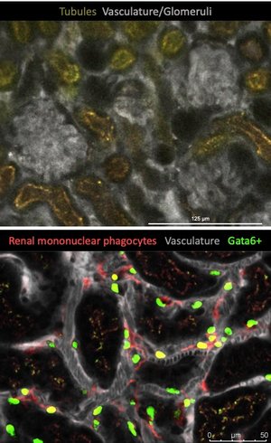
We are also investigating the role of myeloid immune cells in pyelonephritis during the acute phase when the bacteria are translocating from the bladder to the kidneys, but also in the healing phase and the recurrence of pyelonephritis. For these dynamic processes our intravital 2-photon imaging is essential (https://www.baricade.de/).
Another area of interest are S. aureus bloodstream infections and the interaction of these bacteria with Macrophages. On the hand we study why some Kupffer cells, the cells that catch most of S. aureus from the bloodstream, are unable to eliminate the bacteria. Here we are using techniques like dual-RNA sequencing, Flow Cytometry and live cell imaging to reveal the mechanism. In that project we also study the peritoneal cavity and the function of its macrophages. Previous studies of us showed, that the Gata6+ macrophages help to shield S. aureus from neutrophils and delay neutrophil infiltration, which results in more dissemination (Jorch et al., JCI 2019). Now, we have developed a way to visualize the peritoneal cavity with 2-photon microscopy which will help us to gain a deeper view into mechanistically details.
Interested and highly motivated students for lab rotations or master thesis are welcome.
Feel free to contact us and send your CV and possibly a research topic you are interested in to Selina Jorch via Email!